

Congenital anomalies, or malformations of female reproductive tract, are quite common. In a female fetus at its fetal stage, so-called Müllerian ducts must develop and fuse in order to grow uterus, cervix and fallopian tubes. Two paired ducts (Müllerian ducts) develop into female reproductive organs – tubes, uterus, cervix and two-thirds of upper part of vagina. Ovaries and lower third of vagina develop from other structures. If there is a failure in the development or fusion, a female child is born with a malformation. It can cause infertility, diminished fertility, menstrual disorders, multiple miscarriages, premature deliveries or still-born children. Yet they mostly do not cause any problems. 1-3% of all women have a congenital malformation of uterus without even knowing it, unless it affects the fertilisation, embryonic development and delivery.
There are several types of abnormalities of Müllerian ducts:
- Hypoplasia / agenesis – a complete absence of uterus and cervix development (agenesis) or their underdevelopment (hypoplasia). The most common type is Küster-Rokitansky syndrome where uterus and upper part of vagina are underdeveloped or absent. Patients with this syndrome cannot carry a pregnancy, but as they have ovaries these women can have children through surrogacy.
- Uterus unicornis – malformation where uterus is formed from only one Müllerian duct. If one duct is underdeveloped, pregnancy cannot be carried in it. These women also often lack one fallopian tube, but have both ovaries as they do not developed from Müllerian ducts. Yet, in the developed horn of the uterus pregnancy is viable and can be carried out with almost no problems, even though premature births are frequent.
- Double uterus (uterus didelphys, uterus duplex) – a consequence of a complete failure of duct fusion. Each uterine horn is developed and of almost normal size; there are two separate cervices and sometimes a double vagina. Pregnancy can occur in one of the horns and be carried out with no particular problems.
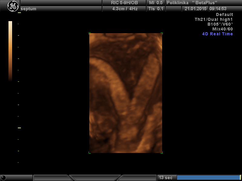
- Heart-shaped uterus (uterus bicornis) – there is a single cervix (also may have a partition), but two horns resulting from incomplete fusion of Müllerian ducts. This shape usually requires surgical correction, which then enables pregnancies with no particular problems.
Septate uterus, arcuate uterus (uterus septus, uterus arcuatus) – the mildest forms of uterine anomalies, an incomplete disappearance of a partition between two horns of uterus. Patients with septate uterus are most frequently associated with reproductive problems, whereas it is still unclear what effects of arcuate uterus on reproduction are.
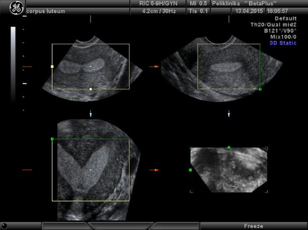
Uterine anomalies are diagnosed with detailed examination and additional methods. 2D ultrasound detects alternations in uterine size and position, and eventual pathological causes such as tumors, cysts, myomas, etc. Nowadays, 3D ultrasound is an indispensable examination for analyzing the structure of genital organs, which is especially important for congenital anomalies. Additional methods that can be used are hysterosalpingography (HSG) with vaginal contrasting, today more with ultrasound (sono HSG) than with X-ray. Hysteroscopy is an endoscopic method that examines the uterus from the inside, it is appropriate for diagnostics and treatment of intrauterine barriers. Laparoscopy is also an endoscopic method for examining abdominal organs with optics. It can detect various anomalies in the uterus, accretions, etc. Treatment for uterine anomalies clearly depends on the cause and clinical findings. In asymptomatic cases, treatment is not necessary.